-
Viewpoint on 'IONS'
Viewpoint on 'Scientific Literacy'
- Proudly sponsored by
-


-
The Dark Side of Lasers

Some scientific discoveries completely change how we look at things: new experiments show that laser theory should be extended to include dark pulse emission.
-
Decoding the Code of Life
Reading information stored by genes — also known as gene sequencing — is a vital task to the study of life itself. A radically new technology has set the stage for a revolution in the deciphering of DNA strands.
-
Honey, I Shrunk the Microscope!
Many of the everyday objects that we use are small enough to fit in our pockets. Take cell phones, with all their different accessories, for instance: are they also likely to come equipped with microscopes the size of a US quarter one day?
Volume 10 Story 5 - 8/9/2010
Cerenkov Photons:
Millions of people fall victim to cancer every year. A great number of these lives could be saved if a simple, inexpensive tool for the early detection of cancer were available. Now, a new technique called Cerenkov luminescence tomography, looks likely to offer a ray of hope in that direction.
 Cancer, tumour, malignancy. If a physician used any of these terms in a diagnosis, it would certainly send a chill down the patients spine. Medical science has made giant strides and developed a plethora of tools to fight cancer. Unfortunately, however, these are only effective if the disease is diagnosed at an early stage. This, then, brings our focus to cancer diagnostics, an area which, despite extensive research, continues to remain quite expensive. Now, a new technique called Cerenkov luminescence tomography, developed by Changqing Li and co-workers at the University of California, Davis, USA could bring this cost down considerably.
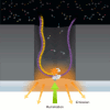
"Cerenkov luminescence is a very interesting phenomenon," notes Li. "When charged particles such as electrons or positrons move faster than the speed of light they emit visible photons." But wait a minute... nothing can travel faster than light, surely? While this is, of course, true, here we are referring to the speed of light in a vacuum. In a given medium, however, light slows down by a factor equal to that mediums refractive index. It is, hence, possible to have particles traveling in a medium with a speed higher than the speed of light in that particular medium. If such particles are charged, they emit visible light, known as Cerenkov luminescence.
Oncologists, the cancer specialists, have primarily relied on biopsies to detect cancer. This technique consists in surgically extracting a small piece of the suspected cancer tissue from the patient, and pathologically examining it. As can be imagined, this cannot be a pleasant experience for the patient. Nowadays, even though the need for biopsies is not completely eliminated, the number of biopsies required on a patient has been significantly reduced thanks to diagnostic tools such as Positron Emission Tomography (PET).
It is well known that cancer tissues have a high metabolic rate. This means they utilize far more glucose than normal tissues to obtain extra energy. In light of this, a good way to detect cancer is to simply check for glucose concentrations in the body, a task at which, in fact, PET does excel.
When using PET, radioactively labeled glucose molecules (Fluorodeoxyglucose or FDG) are injected into the body. These substances decay radioactivelly by emitting positrons, which eventually get annihilated and emit gamma rays. These rays can be detected using an array of gamma ray detectors placed in a ring, which is scanned across the body, thus enabling 3D mapping (tomography) of the labeled molecules. By looking at a PET tomograph, an oncologist can identify tissues suspected to be cancerous.
However, the problem is that very expensive detectors are required in order to detect gamma rays, and this contributes to the ever rising cost of diagnosing cancer. "If we could find a way to use widely available optical detectors, which are cheaper than a typical PET scanner, we could bring down the cost of the system significantly and make cancer diagnosis widely available," points out Li. Instead of detecting the gamma rays, the group has demonstrated that Cerenkov luminescence in the visible spectrum can be detected to achieve results similar to those achieved by PET.
"Cerenkov luminescence is also produced during the radioactive decay of radionuclides (atoms with unstable nuclei and susceptible to radioactive decay) like FDG in biological tissue," says Li. Light travels 25-30% more slowly in biological tissue (with refractive indices of the order of 1.3-1.4) than it does in vacuum (refractive index of 1). The positrons produced by the radioactive decay have energies high enough to travel faster than the slowed down light, giving rise to the emission of visible photons.
The number of visible photons produced depends a great deal on how much faster the positrons travel compared to light. "In our case it is just enough to produce only a handful of photons and this is the major bottle neck," notes Li. "We have overcome this by using a very sensitive camera to efficiently detect the small number of emitted Cerenkov photons. In addition, the procedure is performed in a specially designed light-tight enclosure to exclude ambient light."
The group has demonstrated this technique in mice with tumors. "The procedure involves injection of the radiotracer into the anesthetized animal," describes Li. "The mouse is then placed in the specially designed light-tight box and a very sensitive CCD camera is used to detect non-invasively the faint optical signals that are emitted by the Cerenkov effect and reach the surface of the mouse. Typical imaging time is of a few minutes. A reconstruction algorithm based on a mathemathical model is subsequently employed to estimate the distribution of the optical photons (and therefore the radiotracer that emitted the photons)." This creates the required tomographs (3D images). "The image reconstruction algorithms are very similar to those widely available for bioluminescence optical tomography techniques," adds Li, a fact which reduces further the implementation cost.
It looks increasingly likely that this technique will soon be applied in the diagnosis of patients with cancer. "It can possibly be used to diagnose cancer in any part of the human body that is accessible directly or through the use of endoscopic catheters," explains Li. "This can, for example, be used to detect tumors in the colon." Radiotracers that accumulate in atherosclerotic plaques could also be detected with a catheter-based optical approach. "However, much more technical development will be necessary before this can be made widely available," clarifies Li.
Cerenkov luminescence tomography could possibly go a step further than PET, by acting not just as a diagnostic tool but also by helping in therapeutics. "Cerenkov luminescence tomography can image high energy electrons (beta particles) emitted by radionuclides used and developed for radioimmunotherapy," further clarifies Li. "This is not possible with conventional PET." Radioimmunotherapy is a cancer therapy in which radioactively labeled antibodies that specifically bind to certain cancer tissue are employed. As the antibodies bind, the radiolabel delivers a localized source of toxic radiation to kill the cells. The effectiveness of these therapies depends to a large extend on the precise tracking of the labeled antibodies.
Cerenkov luminescence tomography holds a lot of promise and the scientific community is greatly excited about its applications. "Optical imaging of Cerenkov radiation has emerged as a promising strategy for molecular imaging research as it nicely bridges optical and nuclear imaging," notes Zhen Cheng from the Center for Cancer Early Detection, Stanford University, USA. "Cerenkov luminescence tomography represents another major advancement in the field of Cerenkov optical imaging. This technique could be particularly useful in pre-clinical radiopharmaceutical development and drug discovery. It may also find broad applications in clinical cancer diagnosis."

Cerenkov Photons:
A Cancer Searchlight
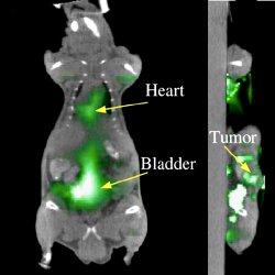
Millions of people fall victim to cancer every year. A great number of these lives could be saved if a simple, inexpensive tool for the early detection of cancer were available. Now, a new technique called Cerenkov luminescence tomography, looks likely to offer a ray of hope in that direction.Tumor imaging by Cerenkov Luminescence Tomography. Reconstructed Cerenkov luminescence tomography images fused with the computed tomography image of mouse. On the left, heart and bladder. On the right, cross-section revealing the presence of a tumor.
"Cerenkov luminescence is a very interesting phenomenon," notes Li. "When charged particles such as electrons or positrons move faster than the speed of light they emit visible photons." But wait a minute... nothing can travel faster than light, surely? While this is, of course, true, here we are referring to the speed of light in a vacuum. In a given medium, however, light slows down by a factor equal to that mediums refractive index. It is, hence, possible to have particles traveling in a medium with a speed higher than the speed of light in that particular medium. If such particles are charged, they emit visible light, known as Cerenkov luminescence.
Oncologists, the cancer specialists, have primarily relied on biopsies to detect cancer. This technique consists in surgically extracting a small piece of the suspected cancer tissue from the patient, and pathologically examining it. As can be imagined, this cannot be a pleasant experience for the patient. Nowadays, even though the need for biopsies is not completely eliminated, the number of biopsies required on a patient has been significantly reduced thanks to diagnostic tools such as Positron Emission Tomography (PET).
It is well known that cancer tissues have a high metabolic rate. This means they utilize far more glucose than normal tissues to obtain extra energy. In light of this, a good way to detect cancer is to simply check for glucose concentrations in the body, a task at which, in fact, PET does excel.
When using PET, radioactively labeled glucose molecules (Fluorodeoxyglucose or FDG) are injected into the body. These substances decay radioactivelly by emitting positrons, which eventually get annihilated and emit gamma rays. These rays can be detected using an array of gamma ray detectors placed in a ring, which is scanned across the body, thus enabling 3D mapping (tomography) of the labeled molecules. By looking at a PET tomograph, an oncologist can identify tissues suspected to be cancerous.
However, the problem is that very expensive detectors are required in order to detect gamma rays, and this contributes to the ever rising cost of diagnosing cancer. "If we could find a way to use widely available optical detectors, which are cheaper than a typical PET scanner, we could bring down the cost of the system significantly and make cancer diagnosis widely available," points out Li. Instead of detecting the gamma rays, the group has demonstrated that Cerenkov luminescence in the visible spectrum can be detected to achieve results similar to those achieved by PET.
"Cerenkov luminescence is also produced during the radioactive decay of radionuclides (atoms with unstable nuclei and susceptible to radioactive decay) like FDG in biological tissue," says Li. Light travels 25-30% more slowly in biological tissue (with refractive indices of the order of 1.3-1.4) than it does in vacuum (refractive index of 1). The positrons produced by the radioactive decay have energies high enough to travel faster than the slowed down light, giving rise to the emission of visible photons.
The number of visible photons produced depends a great deal on how much faster the positrons travel compared to light. "In our case it is just enough to produce only a handful of photons and this is the major bottle neck," notes Li. "We have overcome this by using a very sensitive camera to efficiently detect the small number of emitted Cerenkov photons. In addition, the procedure is performed in a specially designed light-tight enclosure to exclude ambient light."
The group has demonstrated this technique in mice with tumors. "The procedure involves injection of the radiotracer into the anesthetized animal," describes Li. "The mouse is then placed in the specially designed light-tight box and a very sensitive CCD camera is used to detect non-invasively the faint optical signals that are emitted by the Cerenkov effect and reach the surface of the mouse. Typical imaging time is of a few minutes. A reconstruction algorithm based on a mathemathical model is subsequently employed to estimate the distribution of the optical photons (and therefore the radiotracer that emitted the photons)." This creates the required tomographs (3D images). "The image reconstruction algorithms are very similar to those widely available for bioluminescence optical tomography techniques," adds Li, a fact which reduces further the implementation cost.
It looks increasingly likely that this technique will soon be applied in the diagnosis of patients with cancer. "It can possibly be used to diagnose cancer in any part of the human body that is accessible directly or through the use of endoscopic catheters," explains Li. "This can, for example, be used to detect tumors in the colon." Radiotracers that accumulate in atherosclerotic plaques could also be detected with a catheter-based optical approach. "However, much more technical development will be necessary before this can be made widely available," clarifies Li.
Cerenkov luminescence tomography could possibly go a step further than PET, by acting not just as a diagnostic tool but also by helping in therapeutics. "Cerenkov luminescence tomography can image high energy electrons (beta particles) emitted by radionuclides used and developed for radioimmunotherapy," further clarifies Li. "This is not possible with conventional PET." Radioimmunotherapy is a cancer therapy in which radioactively labeled antibodies that specifically bind to certain cancer tissue are employed. As the antibodies bind, the radiolabel delivers a localized source of toxic radiation to kill the cells. The effectiveness of these therapies depends to a large extend on the precise tracking of the labeled antibodies.
Cerenkov luminescence tomography holds a lot of promise and the scientific community is greatly excited about its applications. "Optical imaging of Cerenkov radiation has emerged as a promising strategy for molecular imaging research as it nicely bridges optical and nuclear imaging," notes Zhen Cheng from the Center for Cancer Early Detection, Stanford University, USA. "Cerenkov luminescence tomography represents another major advancement in the field of Cerenkov optical imaging. This technique could be particularly useful in pre-clinical radiopharmaceutical development and drug discovery. It may also find broad applications in clinical cancer diagnosis."
Manoj Mathew
2010 © Optics & Photonics Focus
MM is currently a Technical Manager with the Centre for Cellular and Molecular Platforms (C-CAMP), Bangalore, India.
Changqing Li, Gregory S. Mitchell & Simon R. Cherry, Cerenkov luminescence tomography for small-animal imaging, Optics Letters (2010) 35, 1109-1111 (link).