-
Viewpoint on 'IONS'
Viewpoint on 'Scientific Literacy'
- Proudly sponsored by
-


-
Magnetic Break Up
We usually think of the north and south magnetic poles as an inseparable couple. Experiments have now shown that under the appropriate conditions they can, in fact, break up.
-
Refractive Index: To the Limits and Beyond
Beyond what is naturally possible... metamaterials offer new unexplored opportunities to manipulate light. Researchers show the possibility to enhance the index of refraction of a material beyond natural limits.
-
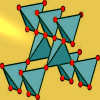
Another Brick in the Nanowall
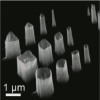
Scientists, just as nano-architects would, are exploring different ways to design nanostructures with fine control over shape and position. A brand-new approach now allows one to build 2D nanowalls up by laying them down brick by brick.
Volume 1 Story 6 - 17/7/2008
The Single Molecule
Who would you hire to localize a single molecule? A new detective is now available! A nano-lightning rod can do the job. It can act as a single-molecule fingerprint detective with an unprecedented spatial resolution up to 15 nanometers.
 When Sherlock Holmes arrives on the scene of a crime, he immediately looks for potential culprits' fingerprints using a magnifying glass. In the same way, scientists can use sharp
metallic nanotips to look for the Raman fingerprints of molecules.
Recently, Jens Steidtner and Bruno Pettinger at the German
Fritz-Haber-Institut of the Max-Planck-Gesellschaft in Berlin have
proven that a metallic nanotip can be employed to localize single
molecules with an unprecedented spatial resolution up to 15
nanometers.
When Sherlock Holmes arrives on the scene of a crime, he immediately looks for potential culprits' fingerprints using a magnifying glass. In the same way, scientists can use sharp
metallic nanotips to look for the Raman fingerprints of molecules.
Recently, Jens Steidtner and Bruno Pettinger at the German
Fritz-Haber-Institut of the Max-Planck-Gesellschaft in Berlin have
proven that a metallic nanotip can be employed to localize single
molecules with an unprecedented spatial resolution up to 15
nanometers.
When light impinges upon a molecule, different parts of the molecule start vibrating at specific frequencies. Just like we all have our characteristic fingerprints, each molecule has its own distinctive set of characteristic frequencies, known as the Raman spectrum of the molecule. These vibrational frequencies have been widely used by scientists as a tool to detect and discriminate molecules, and they are at the core of techniques such as infrared spectroscopy or Raman spectroscopy.
Raman spectroscopy, in particular, has specific advantages that make it a good molecular detective: samples can be prepared easily and it can be used in various environments. However, this detective has some drawbacks that have prevented it from detecting single molecules: poor signal efficiency and a spatial resolution essentially limited to a few hundred nanometers.
Luckily, these disadvantages are overcome by using a more powerful Raman detective, Surface Enhanced Raman Spectroscopy (SERS). In this technique the Raman signal efficiency is greatly enhanced by metal nanostructures brought either in close vicinity to or in contact with the molecule [1]. If the metal nanostructure is a tiny, sharp, and nanosized tip, like a lightning rod at the nanoscale, then the technique is called Tip Enhanced Raman Spectroscopy (TERS). Compared to SERS, TERS is a more efficient molecular detective with spatial resolution at the nanoscale.
"TERS is a very young approach," Pettinger explains, "that combines a raster scanning probe device with a Raman spectrograph. The core of this unit is a tip of suitable material and size that, when illuminated, greatly enhances Raman scattering, making single molecule Raman spectroscopy possible with a spatial resolution around 10-20 nanometers. In a sense, the tip is working as an optical antenna for the incident and radiated light, thereby providing the huge signal enhancement in its close vicinity."
Before Steidtner and Pettinger's work, TERS had only shown indirect evidence for single molecule detection sensitivity. Now, Steidtner and Pettinger have come up with substantial and direct evidence by performing TERS experiments in ultra-high vacuum rather than in air. "One advantage to our approach," Pettinger reveals, "is that the state of the sample surface can be controlled and characterized much better in ultra-high vacuum than in air. In this respect we presented one particularly interesting example: the substantially reduced photobleaching in ultra-high vacuum." Indeed, it is only thanks to this increased stability of the molecule that it has been possible to image single molecules by employing TERS.
This development has significantly improved the spatial resolution of the molecular detective up to 15 nanometers. Renato Zenobi, of the Swiss Federal Institute of Technology (ETH) in Zürich, whose group actively works on TERS technique [2], notes that "the work reveals high sensitivity and localization of TERS at fast signal accumulation times both of which are excellent results."
Experiments like this usually require expertise from different fields, ranging from Raman spectroscopy to scanning probe microscopy, a fact which makes them all the more challenging. According to Pettinger, "the most challenging part was the precise optical alignment in ultra-high vacuum to produce a sharp focus and to get the tip in its center."
When compared to other molecular detectives, "TERS is superior with respect to identification of unknown molecules," Pettinger explains. "It also works for molecules which do not show fluorescence, although the sensitivity drops by about two orders of magnitude." Furthermore, there is more room for improvement in TERS resolution. "The research on TERS started in the year 2000 and," Pettinger remarks, "the quality of the tip is of crucial importance. We routinely get tips with a radius of the apex of about 20 nanometers. Since the achievable resolution depends strongly on this radius, we expect that sharper tips will provide a higher resolution."
"TERS has a wide range of applications," Pettinger concludes, "for example, in surface science, heterogeneous catalysis, single molecule spectroscopy, and biology. So far, these fields have only been briefly explored." Zenobi clarifies that "TERS has immense implications in biology, especially in imaging sub-cellular components such as lipid rafts, membrane protein and others. However, since this work uses ultra-high vacuum, the imaging of bio-entities in this way would be difficult." Although biological entities, like living cells, cannot sustain ultra-high vacuum, this method shows plenty of promise for high resolution chemical imaging which can be further harnessed.
[1] K. Kneipp et al., Surface enhanced Raman scattering: Physics and application, Springer-Verlag, Berlin (2006).
[2] W. Zhang et al., Nanoscale Roughness on Metal Surfaces Can Increase Tip-Enhanced Raman Scattering by an Order of Magnitude, Nano Lett 7, 1401-1405 (2007).

The Single Molecule
Raman Detectives
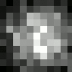
Who would you hire to localize a single molecule? A new detective is now available! A nano-lightning rod can do the job. It can act as a single-molecule fingerprint detective with an unprecedented spatial resolution up to 15 nanometers.A single-molecule fingerprint. TERS image of a single molecule about 10 nm wide.
When light impinges upon a molecule, different parts of the molecule start vibrating at specific frequencies. Just like we all have our characteristic fingerprints, each molecule has its own distinctive set of characteristic frequencies, known as the Raman spectrum of the molecule. These vibrational frequencies have been widely used by scientists as a tool to detect and discriminate molecules, and they are at the core of techniques such as infrared spectroscopy or Raman spectroscopy.
Raman spectroscopy, in particular, has specific advantages that make it a good molecular detective: samples can be prepared easily and it can be used in various environments. However, this detective has some drawbacks that have prevented it from detecting single molecules: poor signal efficiency and a spatial resolution essentially limited to a few hundred nanometers.
Luckily, these disadvantages are overcome by using a more powerful Raman detective, Surface Enhanced Raman Spectroscopy (SERS). In this technique the Raman signal efficiency is greatly enhanced by metal nanostructures brought either in close vicinity to or in contact with the molecule [1]. If the metal nanostructure is a tiny, sharp, and nanosized tip, like a lightning rod at the nanoscale, then the technique is called Tip Enhanced Raman Spectroscopy (TERS). Compared to SERS, TERS is a more efficient molecular detective with spatial resolution at the nanoscale.
"TERS is a very young approach," Pettinger explains, "that combines a raster scanning probe device with a Raman spectrograph. The core of this unit is a tip of suitable material and size that, when illuminated, greatly enhances Raman scattering, making single molecule Raman spectroscopy possible with a spatial resolution around 10-20 nanometers. In a sense, the tip is working as an optical antenna for the incident and radiated light, thereby providing the huge signal enhancement in its close vicinity."
Before Steidtner and Pettinger's work, TERS had only shown indirect evidence for single molecule detection sensitivity. Now, Steidtner and Pettinger have come up with substantial and direct evidence by performing TERS experiments in ultra-high vacuum rather than in air. "One advantage to our approach," Pettinger reveals, "is that the state of the sample surface can be controlled and characterized much better in ultra-high vacuum than in air. In this respect we presented one particularly interesting example: the substantially reduced photobleaching in ultra-high vacuum." Indeed, it is only thanks to this increased stability of the molecule that it has been possible to image single molecules by employing TERS.
This development has significantly improved the spatial resolution of the molecular detective up to 15 nanometers. Renato Zenobi, of the Swiss Federal Institute of Technology (ETH) in Zürich, whose group actively works on TERS technique [2], notes that "the work reveals high sensitivity and localization of TERS at fast signal accumulation times both of which are excellent results."
Experiments like this usually require expertise from different fields, ranging from Raman spectroscopy to scanning probe microscopy, a fact which makes them all the more challenging. According to Pettinger, "the most challenging part was the precise optical alignment in ultra-high vacuum to produce a sharp focus and to get the tip in its center."
When compared to other molecular detectives, "TERS is superior with respect to identification of unknown molecules," Pettinger explains. "It also works for molecules which do not show fluorescence, although the sensitivity drops by about two orders of magnitude." Furthermore, there is more room for improvement in TERS resolution. "The research on TERS started in the year 2000 and," Pettinger remarks, "the quality of the tip is of crucial importance. We routinely get tips with a radius of the apex of about 20 nanometers. Since the achievable resolution depends strongly on this radius, we expect that sharper tips will provide a higher resolution."
"TERS has a wide range of applications," Pettinger concludes, "for example, in surface science, heterogeneous catalysis, single molecule spectroscopy, and biology. So far, these fields have only been briefly explored." Zenobi clarifies that "TERS has immense implications in biology, especially in imaging sub-cellular components such as lipid rafts, membrane protein and others. However, since this work uses ultra-high vacuum, the imaging of bio-entities in this way would be difficult." Although biological entities, like living cells, cannot sustain ultra-high vacuum, this method shows plenty of promise for high resolution chemical imaging which can be further harnessed.
[1] K. Kneipp et al., Surface enhanced Raman scattering: Physics and application, Springer-Verlag, Berlin (2006).
[2] W. Zhang et al., Nanoscale Roughness on Metal Surfaces Can Increase Tip-Enhanced Raman Scattering by an Order of Magnitude, Nano Lett 7, 1401-1405 (2007).
G. V. Pavan Kumar
2008 © Optics & Photonics Focus
PK is currently working as a researcher at ICFO - The Institute of Photonic Sciences, Barcelona (Spain)
J. Steidtner and B. Pettinger, Tip-enhanced Raman spectroscopy and microscopy on single dye molecules with 15 nm resolution, Phys Rev Lett (2008) 100, 236101 (link).